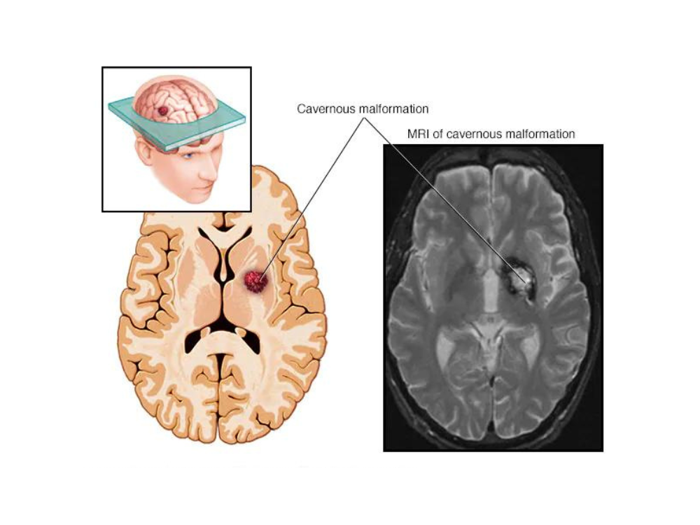

A 33-year-old filmmaker presented with sudden onset of a headache, double vision and MRI brain showed hemorrhage in the midbrain (Upper part of brainstem which connects the brain to the spinal cord). The MRI suggested a Cavernoma ( Vascular Malformation) which had caused the bleed. Since this can bleed repeatedly incapacitating the patient, it was decided to operate and remove the Cavernoma.
The video shows the brains scans before surgery, the surgical approach known as Occipital transtentorial approach. The surgery is done with the patient in sitting position with intraoperative monitoring of III, IV, VI cranial nerves which move the eyes. This monitoring allows and damages to the nerves to be identified early during surgery & hence allow them to be preserved. The midbrain hematoma was reached using neuronavigation and a small cut was made in the midbrain and the Cavernoma was removed. The postop MRI brain showed the Cavernoma was completely removed. The patient recovered her eye functions and is now back to work.
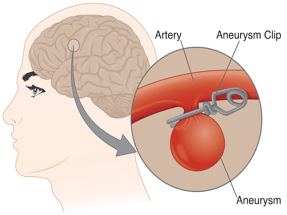
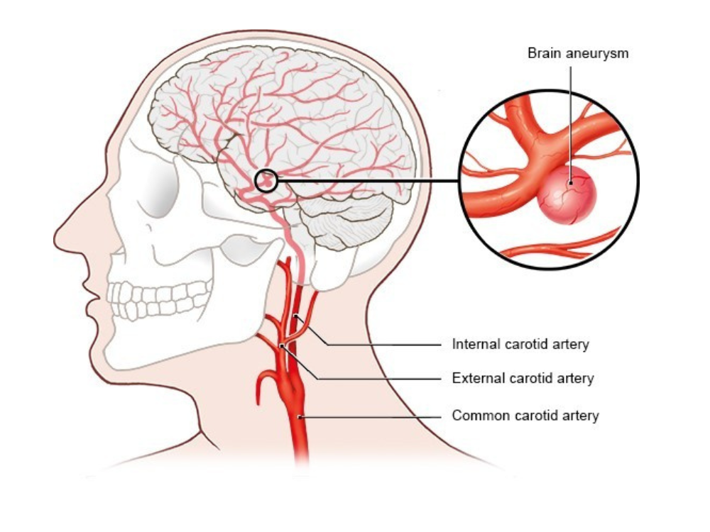
The surgical approach used was right extended fronto-orbitozygomatic, transsylvian, transclinoidal, anterior transpetrosal approach and clipping of the aneurysm.
The surgery video shows the skull base approach made in preparation of clipping of the aneurysm. The anterior petrosal corridor was finally used for temporary control of basilar artery and clipping of the aneurysm. The patient required aVP shunt for hydrocephalus postop and is doing well.

Renowned Neurosurgeon, Cerebrovascular Specialist, and Leader in Advanced Neurosurgical Care. Elevating Lives Through Expertise and Innovation.


Leading Neurological Care Team, Transforming Lives Through Expertise and Compassion.
Copyright © 2025 Neuro Doctors All Rights Reserved.
Designed By Gladias